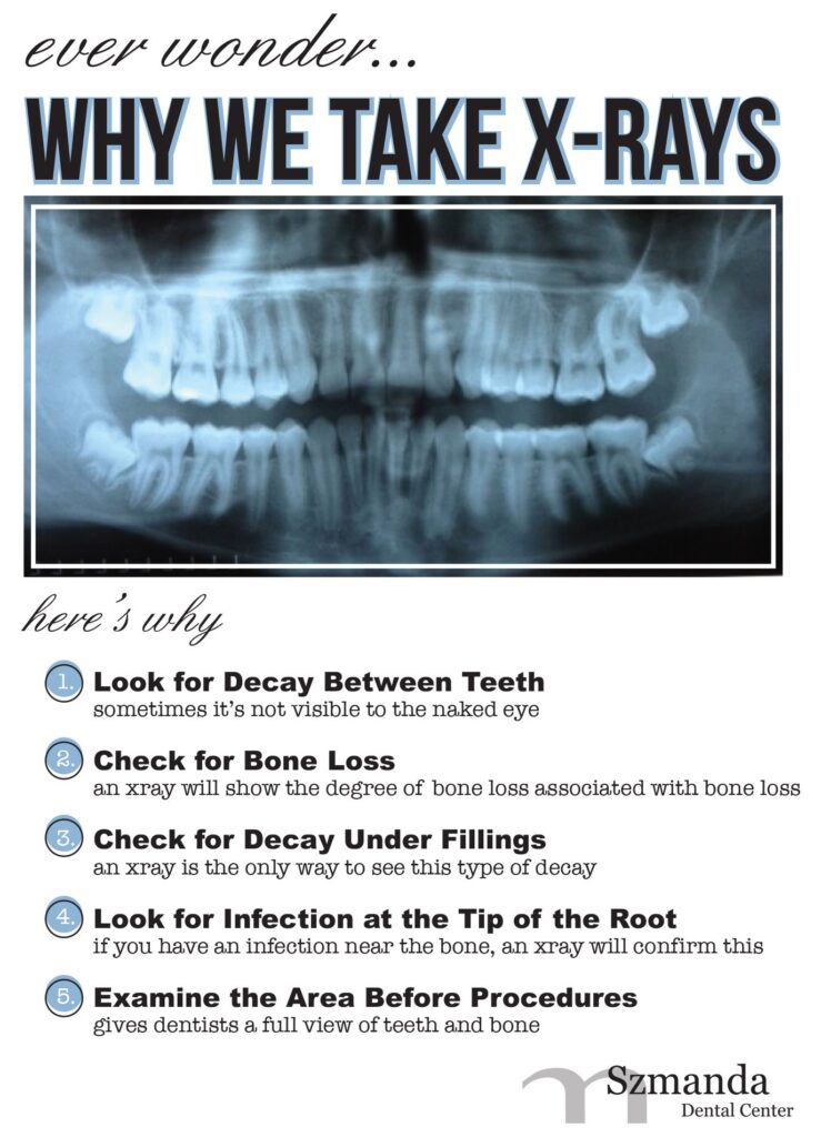
A dental radiograph is sometimes known as a dental x-ray. It is one of the dentist’s most crucial
diagnostic tools since it provides a clearer image of your teeth’s condition than a simple oral
examination.
The Bitewing X-Ray
Because they are a fantastic way to spot any decay between teeth or below the gumline,
bitewing X-rays are extremely popular and frequently used for prophylactic purposes. Because
patients must bite down on the X-ray film, the term “bitewing” was coined. These X-rays can be
taken while the patient is sitting in the dentist chair.
Precision X-Ray
Bitewings reveal the majority of the tooth, but a periapical X-Ray is preferable if your dentist
needs to see your complete tooth or jawbone. An image of the complete tooth, including a little
portion of the tooth root, is captured by this kind of X-ray.
The complete upper or lower row of teeth is often shown in one image on the X-ray. If your
dentist suspects difficulties with the jawbone or damage to the tip of the tooth root, these kinds
of X-rays could be taken.
Occlusal X-Ray
Occlusal X-rays are made to record what occurs inside the roof or floor of the mouth, allowing
the dentist to see the whole development and positioning of the teeth. This may be used to
identify supernumerary (additional) teeth that could harm good permanent teeth or to determine
why teeth haven’t yet erupted.
Panoramic X-Ray
Panorama x-rays are similar to panorama pictures. Instead than merely a “snapshot” of a few
teeth, they display the entirety of the mouth. These images are acquired from the outside of the
mouth as opposed to the inside, unlike regular dental x-rays.
The patient does not move as an x-ray machine circles their head in an arc from one side to the
other. The end result is a single, extended image that displays every tooth and jaw as well as
the nasal passage and sinuses.
Dental X-Rays in Milpitas

